
Page 3 - HarmfulAlgaeReadMe
P. 3
Methods and Evaluation of Data
Quality
Surface seawater samples for phytoplankton analysis
are collected with a Niskin bottle mounted on a pole (or
directly by submerging the sample container) at each
station, with additional water samples from 5, 10, and
20 m depths being collected at one station per patrol in
the same manner as the nutrient samples. Once
collected, samples are immediately preserved by
adding Lugol’s iodine solution (final concentration in the
sample 1–2%).
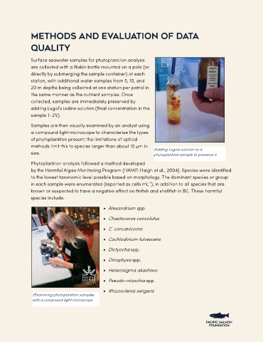
Samples are then visually examined by an analyst using
a compound light microscope to characterize the types
of phytoplankton present; the limitations of optical
methods limit this to species larger than about 10 µm in
1Adding Lugols solution to a
size. phytoplankton sample to preserve it.
Phytoplankton analysis followed a method developed
by the Harmful Algae Monitoring Program (HAMP, Haigh et al., 2004). Species were identified
to the lowest taxonomic level possible based on morphology. The dominant species or group
-1
in each sample were enumerated (reported as cells mL ), in addition to all species that are
known or suspected to have a negative effect on finfish and shellfish in BC. These harmful
species include:
• Alexandrium spp.
• Chaetoceros convolutus
• C. concavicorne
• Cochlodinium fulvescens
• Dictyocha spp.
• Dinophysis spp.
• Heterosigma akashiwo
• Pseudo-nitzschia spp.
• Rhizosolenia setigera
2Examining phytoplankton samples
with a compound light microscope